Verwendung: Dieses Produkt ist ausschließlich für Forschungszwecke bestimmt.
Alle Produktinformationen auf dieser Website dienen ausschließlich Bildungszwecken. Jede Form der Einbringung in den Körper von Menschen oder Tieren ist gesetzlich streng verboten. Dieses Produkt darf nur von zugelassenen und qualifizierten Fachleuten gehandhabt werden. Es ist kein Arzneimittel, Lebensmittel oder Kosmetikum und darf nicht als solches gekennzeichnet, verwendet oder falsch etikettiert werden.
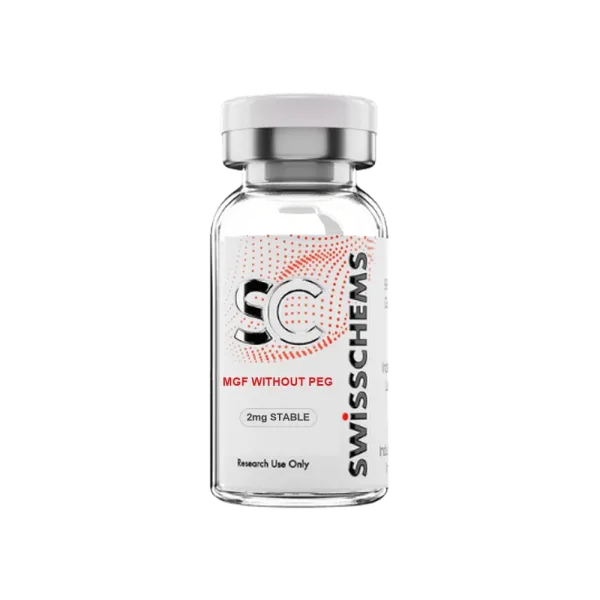
MGF without PEG 2mg
Während der Umstellung unserer Verpackung erhalten Sie möglicherweise Produkte mit dem bisherigen oder dem neuen Etikett. Formulierung, Reinheit und Qualität bleiben nach unseren Standards genau gleich.
- Kreditkarte
- Banküberweisung (SEPA Echtzeit)
- Banküberweisung (ACH)
- Wise
- Revolut
- Zelle
- Remitly
- Western Union
- Chime
- Andere
-25% auf alles – bis 31. Oktober
25% auf reguläre Preise statt anderer Angebote. Nur mit dem Krypto-Zahlungsrabatt kombinierbar.
Jede Charge wird auf Reinheit, Wirksamkeit und Qualität geprüft.
Laborergebnisse ansehen →Mechano Growth Factor (MGF) is the alternative splice variant of the insulin-like growth factor 1 (IGF-1) gene, also designated IGF-1Ec, distinguished from the systemic IGF-1Ea isoform by a distinct C-terminal E-domain peptide sequence.
Research interest centers on its proposed role in activating satellite cell proliferation in skeletal muscle tissue following mechanical loading, distinct from the growth-promoting signalling of mature IGF-1. Research applications include satellite cell activation pathway studies, IGF-1 splice variant comparative research, and muscle tissue mechanotransduction investigation.
- High Purity – 99% Purity Guaranteed
- Independently Lab Tested
- Research Grade Quality
- For Laboratory Research Use Only
| Chemical Formula | C121H200N42O39 |
|---|---|
| Synonyms | MGF, IGF-1Ec |
| Molar Mass | 2,644.85 g/mol |
| CAS Number | 1565841-06-6 |
| Total Compound Content | 2mg per vial |
| Shelf Life | 36 months |
Every batch is independently lab tested for identity, purity and potency. View our lab testing program →
How does MGF (IGF-1Ec) functionally differ from mature IGF-1 (IGF-1Ea) in muscle tissue research models?
Both are products of the same IGF-1 gene but result from alternative splicing, producing distinct C-terminal E-domain peptide sequences with reportedly distinct biological roles. Mature IGF-1 (IGF-1Ea) primarily promotes protein synthesis and hypertrophy in already-differentiated myoblasts via PI3K/Akt/mTOR signalling.
MGF (IGF-1Ec) is studied for a proposed earlier-stage role: activating quiescent satellite cells to re-enter the cell cycle and begin proliferating, a necessary precursor step before these cells can differentiate and contribute to muscle fiber repair or growth.
Research distinguishing these roles typically uses satellite cell proliferation assays (Ki-67, BrdU incorporation) for MGF versus protein synthesis assays (puromycin incorporation, mTOR pathway activation) for mature IGF-1.
What is the significance of using non-PEGylated MGF in pharmacokinetic and mechanistic research?
PEGylation (covalent attachment of polyethylene glycol chains) is a common modification used to extend a peptide's circulating half-life by reducing renal clearance and proteolytic degradation.
Non-PEGylated MGF reflects the native peptide's pharmacokinetic profile, which is relevant for research questions about the compound's natural biological half-life and clearance mechanisms, or where the larger PEG moiety might sterically interfere with receptor binding or cellular uptake in a specific experimental model.
Researchers should select PEGylated or non-PEGylated formats based on whether their study question concerns native peptide kinetics or extended-exposure pharmacology.
What experimental models are used to study MGF's proposed satellite cell activation mechanism?
Isolated satellite cell cultures (from skeletal muscle biopsy or whole-muscle explant) and ex vivo muscle fiber preparations are standard models for studying MGF's effects on satellite cell quiescence exit and proliferation, typically using cell-cycle markers (Ki-67, PCNA) and proliferation assays (BrdU or EdU incorporation) as primary readouts. Mechanical loading or injury models (such as eccentric contraction protocols in ex vivo muscle preparations) are sometimes used to study endogenous MGF splice variant induction alongside exogenous MGF administration, allowing comparison of native versus exogenous peptide effects on the same satellite cell population.